No hay productos en el carrito.
Algunos apuntes básicos…
La vascularización de la rodilla corre a cargo de ramas de la arteria poplítea, esta arteria, junto con la vena poplítea, es continuación de la arteria y vena femoral y se hacen posteriores al pasar por debajo del hiato del aductor (hiato que se forma por la inserción más distal del músculo aductor mayor en el tubérculo del aductor, relieve óseo superior al cóndilo medial femoral).

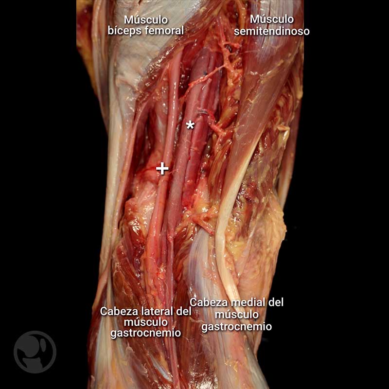
Visión posterior de la rodilla o región poplítea donde se visualiza la arteria y vena poplítea (*) responsable de la vascularización de la rodilla, acompañados por el nervio tibial (+). Esta zona posterior esta limitada por unos músculos especificados en la imagen y que marcan los límites del rombo poplíteo.
En esta zona posteromedial, estos vasos dan una serie de ramas que con un trayecto horizontal se dirigen de posterior a anterior con la finalidad de vascularizar toda la rodilla. Estas arterias se denominan, según la nómina anatómica, arterias articulares de la rodilla (Feneis, 2000), sin embargo en los libros clásicos de anatomía y en muchos artículos científicos se continúan nombrando arterias geniculares de la rodilla (Teixeiro de Carvalho, 2016).
Como el segundo término es más frecuente y habitual nos referiremos a ellas como arterias y venas geniculares. Estos vasos son 5, la arteria genicular media, la superolateral, superomedial, la inferolateral y inferomedial.

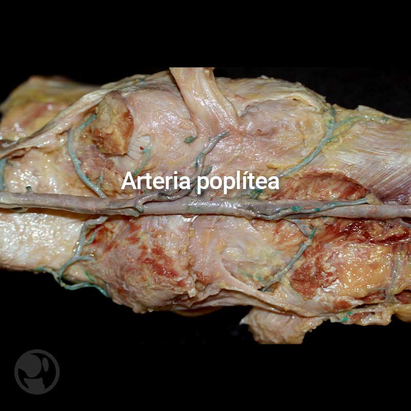
Imagen de una arteria poplítea inyectada donde se visualizan todas las arterias geniculares y algunas ramas musculares cortadas.

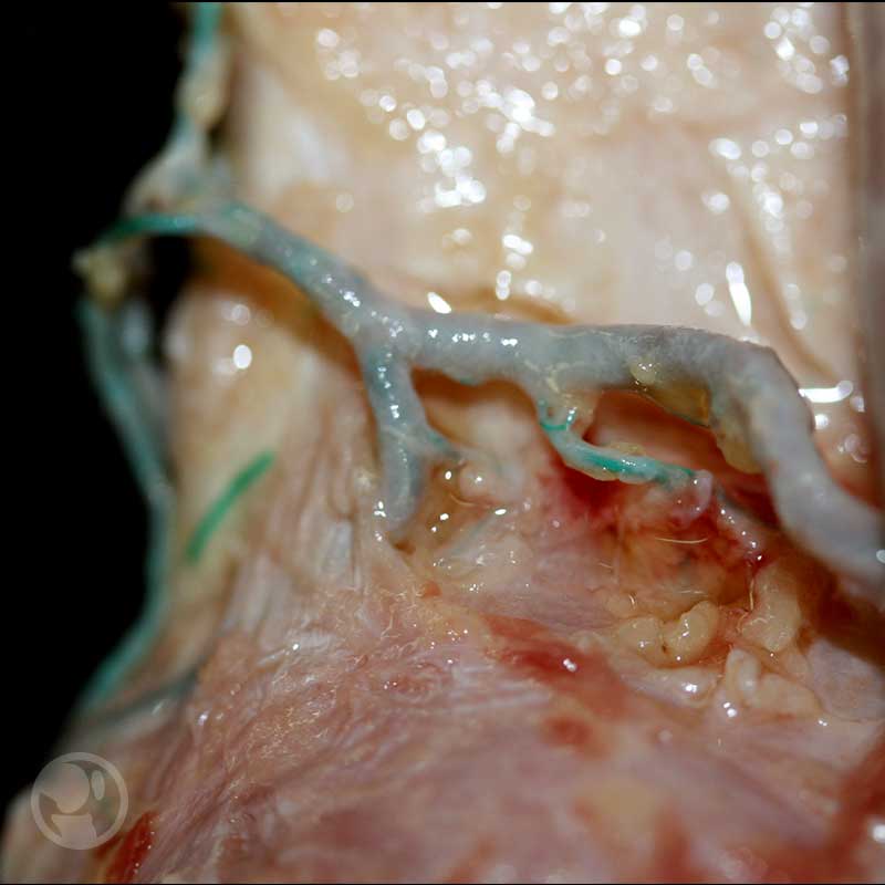
La imagen de menor tamaño, muestra como una de estas arterias articulares pueden quedar excesivamente fijadas por la presencia de un ligamento no constante aunque si presente con relativa frecuencia (*)
Knee Meniscos, Cruzados y Aparato Extensor
Curson online
(Pulsa la foto para acceder)
“Knee meniscos, cruzados y aparato extensor” te permitirá diagnosticar y tratar de manera efectiva y clara la patología más frecuentes de la rodilla; una vez finalices este curso serás capaz de diagnosticar, tratar y seguir lesiones de: meniscos, cruzado anterior, posterior y aparato extensor. También estarás en capacidad de ofrecer opciones terapéuticas médicas, ortopédicas y quirúrgicas para cada una de estas patologías.
Arteria genicular media
Viene por la cara anterior de la arteria poplítea y con un corto trayecto hacia la parte posterior de la cápsula articular, entra proximal al ligamento poplíteo oblicuo, entre los dos cóndilos lateral y medial. Esta arteria es la responsable de la vascularización de los ligamentos cruzados y el tejido sinovial. Pero también es muy susceptible de ser lesionada en cirugía y traumatismos de la rodilla por ello tal como describen algunos autores (Shahid et cols, 2015).
Arteria genicular superomedial
Se dirige al músculo vasto medial pasando por delante del tendón del músculo aductor mayor.
Arteria genicular superolateral
Se dirige al músculo vasto lateral, pasando anterior al músculo bíceps femoral.
Arteria genicular inferomedial
Recorre el borde proximal del poplíteo y se dirige a anterior entre la tibia y el ligamento colateral medial.
Arteria genicular inferolateral
Cruza al poplíteo y pasa profundamente al ligamento colateral lateral. Estos vasos atraviesan diferentes estructuras desde su origen en la arteria poplítea pero están más o menos libres en ese punto de origen posterior.
Sin embargo, hay variaciones descritas (Miguel y cols. 2006) en las que se observa un ligamento que fija alguna de estos vasos geniculares (generalmente el superolateral) y ello pueda favorecer lesiones de las mismas cuando se manipulan los vasos poplíteos en cirugía posterior de la rodilla. Todos estos vasos hacen anastomosis en la zona anterior de la rodilla y entorno a la articulación de la rodilla, junto con otras arterias como son la arteria descendente de la rodilla (rama de la arteria femoral que viene con el nervio safeno por el conducto del adductor) y ramas recurrentes de la arteria tibial y peronea.
Imagen del banner por Nino Liverani on Unsplash
- Bibliografía:
1.- Ryu K, Iriuchishima T, Oshida M, Saito A, Kato Y, Tokuhashi Y, Aizawa S. Evaluation of the morphological variations of the meniscus: a cadaver study. Knee Surg Sports Traumatol Arthrosc. 2015; 23:15-9.
2.- Bozkurt M, Elhan A, Tekdemir I, Tönük E. An anatomical study of the meniscofibular ligament. Knee Surg Sports Traumatol Arthrosc. 2004; 12: 429-33.
3.- Tajima G, Nozaki M, Iriuchishima T, Ingham SJ, Shen W, Smolinski P, Fu FH. Morphology of the tibial insertion of the posterior cruciate ligament. J Bone Joint Surg Am. 2009; 91: 859-66.
4.- Brooker B, Morris H, Brukner P, Mazen F, Bunn J. The macroscopic arthroscopic anatomy of the infrapatellar fat pad. Arthroscopy. 2009; 25: 839-45.
5.- Dombrowski ME, Costello JM, Ohashi B, Murawski CD, Rothrauff BB, Arilla FV, Friel NA, Fu FH, Debski RE, Musahl V. Macroscopic anatomical, histological and magnètic resonance imaging correlation of the lateral capsule of the knee. Knee Surg Sports Traumatol Arthrosc. 2016; 24: 2854-60.
6.- Claes S, Vereecke E, Maes M, Victor J, Verdonk P, Bellemans J. Anatomy of the anterolateral ligament of the knee. J Anat. 2013; 223: 321-8.
7.- Irvine GB, Dias JJ, Finlay DB. Segond fractures of the lateral tibial condyle: brief report. J Bone Joint Surg Br. 1987; 69: 613-4.
8.- Hughston JC, Andrews JR, Cross MJ, Moschi A. Classification of knee ligament instabilities. Part II. The lateral compartment. J Bone Joint Surg Am. 1976; 58:173-9.
9.- Haims AH1, Medvecky MJ, Pavlovich R Jr, Katz LD. MR imaging of the anatomy of and injuries to the lateral and posterolateral aspects of the knee. AJR Am J Roentgenol. 2003; 180: 647-53.
10.- Moorman CT, LaPrade RF. Anatomy and biomechanics of the posterolateral corner of the knee. J Knee Surg. 2005; 18:137-45.
11.- Campos JC, Chung CB, Lektrakul N, Pedowitz R, Trudell D, Yu J, Resnick D. Pathogenesis of the Segond fracture: anatomic and MR imaging evidence of an iliotibial tract or anterior oblique band avulsion. Radiology. 2001; 219: 381-6.
12.- Vincent JP, Magnussen RA, Gezmez F, Uguen A, Jacobi M, Weppe F, Al-Saati MF, Lustig S, Demey G, Servien E, Neyret P. The anterolateral ligament of the human knee: an anatomic and histologic study. Knee Surg Sports Traumatol Arthrosc 2012; 20: 147–152.
13.- De Maeseneer M, Shahabpour M, Lenchik L, Milants A, De Ridder F, De Mey J, Cattrysse E. Distal insertions of the semimembranosus tendon: MR imaging with anatomic correlation. Skeletal Radiol. 2014 ;43: 781-91.
14.- Aragão JA, Reis FP, de Vasconcelos DP, Feitosa VL, Nunes MA. Metric measurements and attachment levels of the medial patellofemoral ligament: an anatomical study in cadavers. Clinics (Sao Paulo). 2008; 63: 541-4.
15.- Reider B, Marshall JL, Koslin B, Ring B, Girgis FG. The anterior aspect of the knee joint. J Bone Joint Surg Am. 1981;63: 351-6.
16.- Feneis H. Nomenclatura anatómica ilustrada. 4th Edición. Barcelona. Editorial Masson. 2000.
17.- Texeira de Carvalho RT, Ramos LA, Novaretti JV, Ribeiro LM, Szeles PR, Ingham SJ, Abdalla RJ Relationship Between the Middle Genicular Artery and the Posterior Structures of the Knee: A Cadaveric Study. Orthop J Sports Med. 2016; 9: 4-12.
18.- Shahid S, Saghir N, Cawley O, Saujani S. A cadaveric study of the branching pattern and diameter of the genicular arteries: a focus on the middle genicular artery. J Knee Surg. 2015; 28: 417-24.
19.- Miguel M, Ortiz JC, Calzada J, Llusà M, Lorente M, Götzens V. An inconstant ligament in the popliteal region associated to the superior genicular arteries: surgical importance. Surg Radiol Anat. 2006 ;28: 457-61.
20.- Orduña Valls JM, Vallejo R, López Pais P, Soto E, Torres Rodríguez D, Cedeño DL, Tornero Tornero C, Quintáns Rodríguez M, Baluja González A, Álvarez Escudero J. Anatomic and ultrasonographic evaluation of the knee sensory innervation: A cadaveric study to determine anatomic targets in the treatment of chronic knee pain. Reg Anesth Pain Med. 2017 ;42 :90-98.
21.- Romanes G.J. Cunningham Tratado de Anatomía. 12th edición. Madrid. Editorial interamericana_McGraw-Hill. 1987.
22.- Nordin M, Frankel VH. Basic biomechanics of the musculoskeletal system.
23.- Gongora LH, Rosales CM. Articulación de la rodila y su mecánica articular. Medisan 2003; 7 (2)


